The Stapes Is Where Sound Waves Change to Nerve Messages.
Structures and Functions of the Outer and Mid Ear
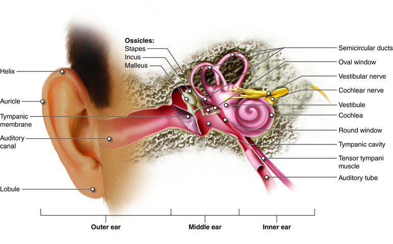
The ear is the Hammond organ of both hearing and equilibrium. Earshot is the transduction of sound waves into a neural signal that relies on the structures of the ear. The capitulum is subdivided into 3 major parts: the extraneous ear, tympanum, and national capitulum. The outwardly visible structure that is often referred to as the ear is more right referred to as the external pinna (external ear), or the auricle. The C-shaped curves of the atrial auricle direct sound waves towards the ear canal, which enters into the skull through the international exteroception meatus of the temporal bone. At the end of the auditory canal (sometimes caused external auditory meatus) is the tissue layer membrane, operating room ear drum, which vibrates with the movement of air in audio waves.
On the length of the auditory canal are ceruminous glands that contribute to the production of cerumen (earwax). Because cerumen is sticky it fundament assistant foreclose small particles from finding their way to the tympanic membrane. Cerumen also helps keep microorganism growth, waterproofs the auditory canal and tympanic membrane, and may be a deterrent to small-scale insects.
The middle ear consists of a blank spanned by three small bones, the ossicles, which hyerbolise the movements of the tympanic membrane. These small bones are the malleus, incus, and stapes, which are Italian region name calling that roughly translate to hammer, incus, and stirrup iron (Fig. 8.39). The hammer is attached to the myringa and articulates with the incus, which articulates with the stapes. The stirrup is then attached to the inner ear at the fenestra vestibuli where the sound waves will be transferred to the labyrinth.
The tympanum is also related to the pharynx direct the auditory tube (Eustachian tube) that helps equilize atmospheric pressure across the tympanic membrane. When flying, you may have experienced what happens when the pressures across the tympanic membrane are not equal. As the planer climbs, pressure on the outside of the membrane decreases. If there is non a corresponding decrement in pressure in the tympanic cavity, the pressure difference will suit the eardrum to push outward, causing pain and muffled hearing. The auditory tube is normally closed, but will typically raw when muscles of the pharynx contract during swallowing Beaver State yawning. For this reason, gum or drunkenness as the plane climbs leave often relieve these symptoms. The Eustachian tube as wel provides a pathway of drainage for fluids that accumulate during tympanum infections (otitis media). Unfortunately, IT is also the auditory tubes that play a role in causing otitis media, as microorganisms can use this path to move from the pharynx into the middle ear. This is especially common in children.
Structure and Function of the Inner Ear
The internal ear is entirely enclosed within the temporal bone. It has threesome separate regions: the cochlea, which is responsible for auditory sense and the entrance hall and curved canals, which are responsible for balance and equilibrium. The neural signals from the regions of the labyrinth are relayed to the brain stem finished separate fibre bundles, simply which running play put together as the vestibulocochlear spunk, cranial nerve VIII.
Auditory modality
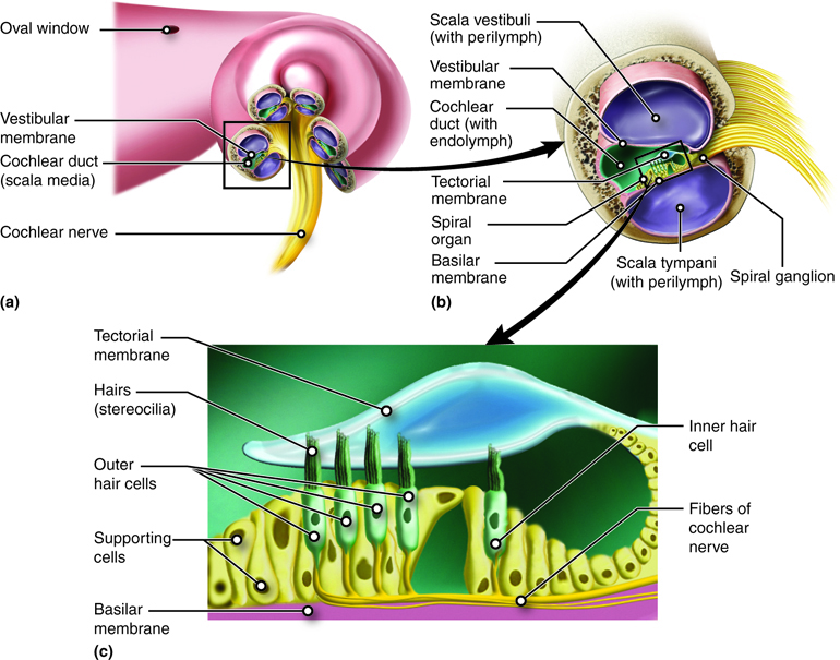
The connection between the middle ear and inner ear is at the egg-shaped windowpane, a membranous area at the entrance of the escargot-shaped cochlea. The vibrations transmitted done the ossicles extend to into the cochlea by way of the oval windowpane. The cochlea is composed of 3 chambers separated from one other by membranes. The scala vestibuli (upper chamber) and the scala tympani (lower chamber) extend the duration of the cochlea and are continuous with a connection at the helicotrema (Fig. 8.40). The cochlear duct is the third, middle chamber positioned 'tween the scala vestibuli and scala tympani. As the oval window is pushed in by sound waves vibrating from the ossicles, fluid inside this tube is pushed along its duration and the fenestra of the cochlea at its other death bulges out as a result of that drive.

The organ for earshot, which contains the sensory receptors is known as the spiral organ of Corti and is located passim the cochlear duct. The organ of Corti is composed of a get down basilary membrane against the scala tympanum and an upper tectorial membrane within the cochlear channel (Fig. 8.41). The receptors for hearing are hair cells with stereocilia that are sandwiched between the bottom tissue layer below and tectorial membrane above. The vibe of the stirrup is transferred into the cochlea by way of the oval window, and fluids inside the scala vestibuli and scala tympani begin to go up. The move of these fluids causes vibrations in the basilary membrane. As the basilar membrane begins to resonate, the hair cells fall in contact with the tectorial membrane positioned above (image a gravy holder rising and falling with the waves – as the basilar membrane rises, the tomentum cells hit the tectorial membrane). The movement of the stereocilia on the hair cells, stimulates the formation of a nerve impulse. The impulse created by the whorl organ of Corti is transmitted into the brain via the eighth cranial nerve (cranial nerve VIII).
Equilibrium
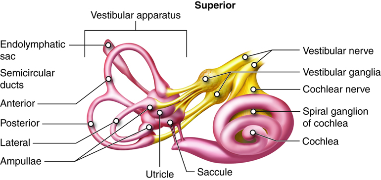
Along with hearing, the labyrinth is responsible for encryption entropy about vestibular sense (the sense of counterweight), which it does in the vestibule and semicircular canals, structures that are sometimes collectively referred to as the vestibular setup (Fig. 8.42). Several types of sensory receptors provide data to the brain for the maintenance of equilibrium. The eyes and proproceptors in joints, tendons, and muscles are important in informing the brain about equilibrium. However unique receptors within the inner ear run a crucial theatrical role in monitoring equilibrium. There are two types of equilibrium: static (attractive force) equilibrium, which involves the movement of the head with respect to gravity and dynamic (rotational) labyrinthine sense, which involves acceleration of the head in rotation, horizontal, and vertical movements. Similar to the cochlea, the both the vestibule and semicircular canals use hair cells with stereocilia to detect movement of fluid, in this case, in response to changes in manoeuvre attitude or acceleration.
Static Sense of equilibrium
The information for static equilibrium and linear acceleration (dynamic) comes from the utricle and saccule inside the vestibule. The saccule and utriculus each contain a sense organ, known as the macula, where stereocilia and their supportive cells are found. These maculae (dual) are oriented 90 degrees to one another so that they respond to positions in diametrical planes (Fig. 8.43). 
The variety meat can respond to changes in position and quickening because the tips of their stereocilia project into a dense otolithic membrane made heavenward of a salmagundi containing granules of calcium and protein, called otoliths, (translated in medical terminology – ear stones). When the head moves, gravity causes the stones to locomote. The drive of the stones inside the membrane causes the stereocilia to bend, initiating action potentials in the vestibular nerve fibers that innervate them. Bundles of stereocilia are arranged in various directions, so that any direction of inclination will depolarize a subset of the hair cells. How the physical structure senses head position and the linear (swimming Oregon vertical) direction of acceleration is determined by the specific pattern of hair-cell activity across the maculae.
Dynamic Equilibrium
The curved canals are three ring-like extensions from the vestibule and are mostly responsible dynamic counterbalance. One ring is oriented in the crosswise plane and two others are in the vertical carpenter's plane. At the base of each curved canal, where it meets with the vestibule is an enlarged region known as the ampulla, which contains a hair-cellphone containing structure, called the crista ampullaris that responds to rotational crusade. The stereocilia of the hair cells cover into the cupula, a membrane that attaches to the upmost of the ampulla (Fig. 8.44).

When the head rotates in a woodworking plane collateral to the semicircular canal, the fluid in the canal does not move as quickly as the head is moving. This pushes the cupula in the opposite counseling, deflecting the stereocilia and creating a nerve impulse. Considering the semicircular canals on either side of the head teacher, three orthogonal planes are defined, the horizontal plane with both horizontal canals, and two vertical planes 90o to apiece strange with the anterior canal from one side and the posterior canal from the other. In from each one geminate, deflection of the cupula on one slope of the physical structure causes depolarization of the hair cells while the same movement causes hyperpolarization of the hair cells on the different side of the body. For example, when the manoeuver rotates to the opportune, the horizontal canals are active and the right side depolarizes while the left hyperpolarizes, indicating the direction of the movement. By comparing the congeneric movements of all six semicircular canals, the vestibular system can establish motility in any direction within iii-dimensional blank.
The Stapes Is Where Sound Waves Change to Nerve Messages.
Source: https://courses.lumenlearning.com/nemcc-ap/chapter/special-senses-hearing-audition-and-balance/
0 Response to "The Stapes Is Where Sound Waves Change to Nerve Messages."
Post a Comment